WORK 01
Knee Model
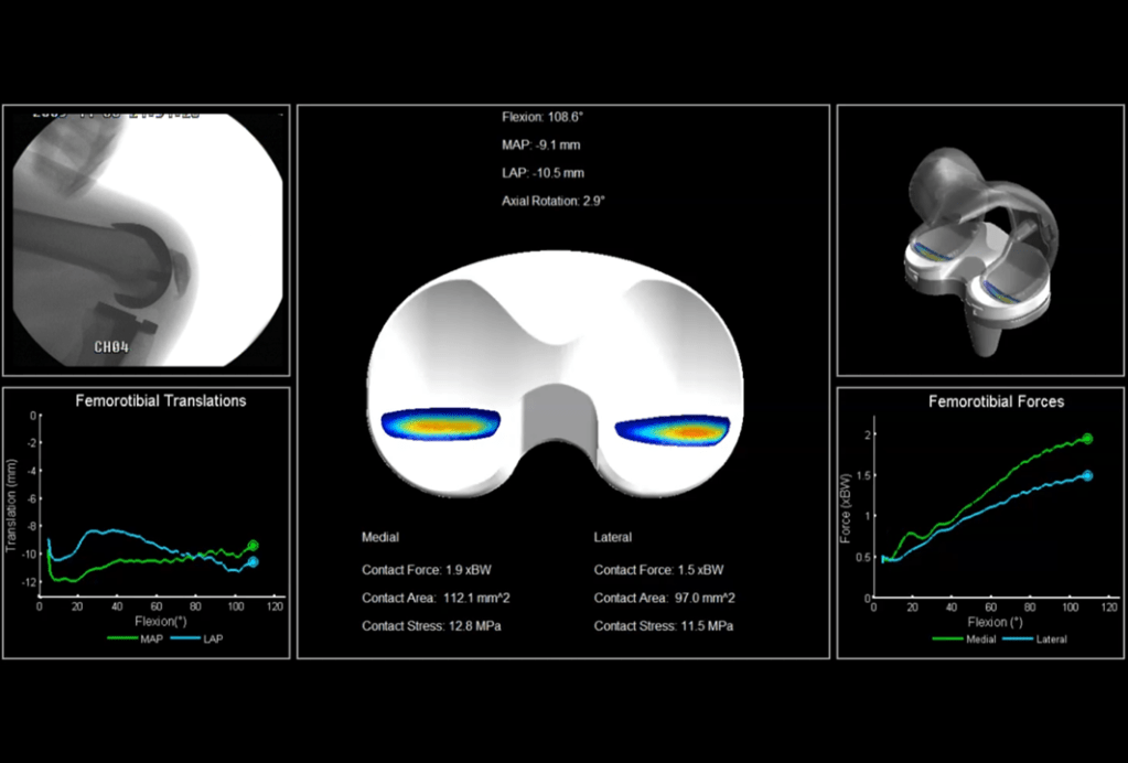
Using mathematical knee modeling provides a valuable approach for assessing the kinematics and kinetics of implanted knees. This technique involves creating computational models that simulate the biomechanical behavior of the knee joint with implants. By inputting various parameters and conditions, researchers can predict how the implanted knee will move and respond during different activities. Mathematical knee modeling offers insights into factors such as range of motion, joint forces, stresses, and alignment, aiding in the optimization of implant design and surgical techniques for improved patient outcomes and long-term functionality.
WORK 02
Hip Model
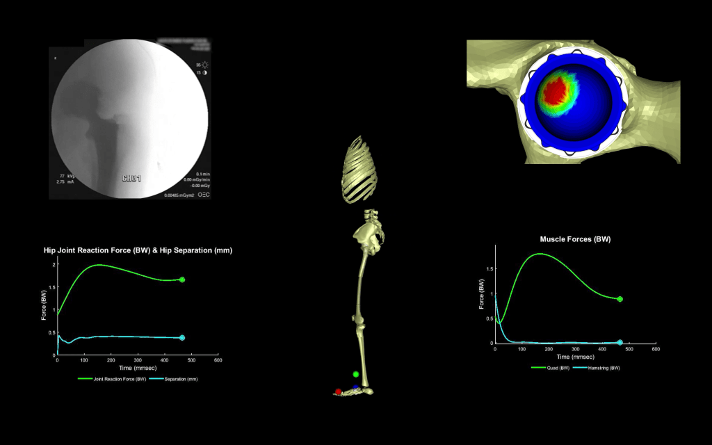
Total hip arthroplasty generally improves patient quality of life but can still cause issues like atypical forces, premature component wear, and abnormal kinematics compared to natural joints. Common complications include instability, separation, sliding, and edge loading within the hip joint. Evaluating potential solutions for these issues is costly and time-consuming. However, mathematical modeling offers an accurate and efficient way to explore solutions. The study aims to develop and validate a mathematical model to compare different hip implant designs and identify factors contributing to hip separation, instability, and edge loading. The model has been validated against data and shows that shifting the joint rotation center during surgery may lead to postoperative instability, emphasizing the need for precise implantation to reduce risks.
WORK 03
Machine Learning
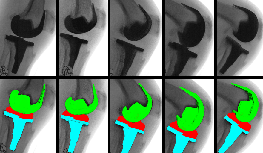
Leveraging machine learning techniques for image segmentation and image registration in implanted knees holds significant promise. By training algorithms on large datasets of medical images, machine learning can effectively identify and separate different anatomical structures in images, enabling precise segmentation of bone, implant components, and surrounding tissues. Moreover, machine learning-driven image registration can align pre- and post-operative images, allowing for accurate comparisons and assessments of implant placement, alignment, and changes over time. This innovative approach enhances clinicians’ ability to monitor the condition of implanted knees, optimize surgical procedures, and ultimately contribute to improved patient care and outcomes.
WORK 04
Fluoroscopy Evaluation
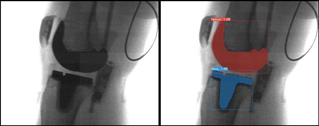
Researchers employed video fluoroscopy to examine the femorotibial contact patterns in subjects who underwent either posterior cruciate retention or posterior cruciate substitution total knee arthroplasty. They analyzed the femorotibial contact of many individuals with different types of total knee replacements performed by various surgeons. Using successive deep knee bends at different angles, video images were captured and processed.